History of Present Illness
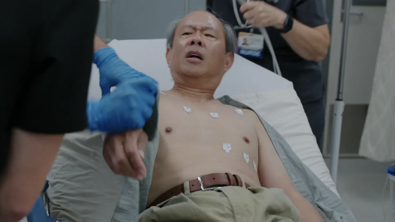
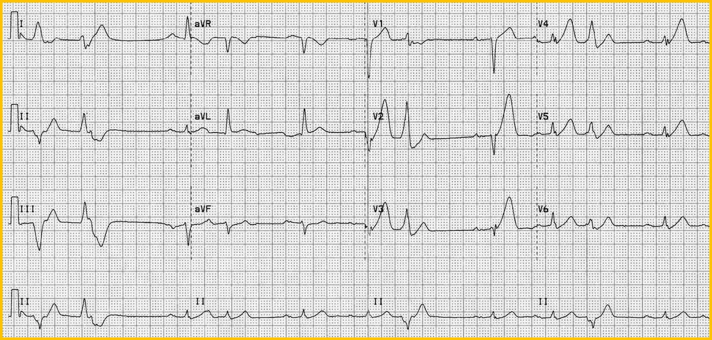
Adult male brought to the ED by a coworker after about 10 minutes of substernal chest pain with diaphoresis. Triage EKG showed an anterior STEMI and a Code STEMI was called to Trauma 2. He describes the pain as 'like an elephant is on my chest' and has never had a heart attack before. patient has no known allergies and is taking metformin and rosuvastatin implying type 2 diabetes or prediabetes and hyperlipidemia. Triage blood pressure was 170/92.
Emergency Department Course
Anterior STEMI Arrival
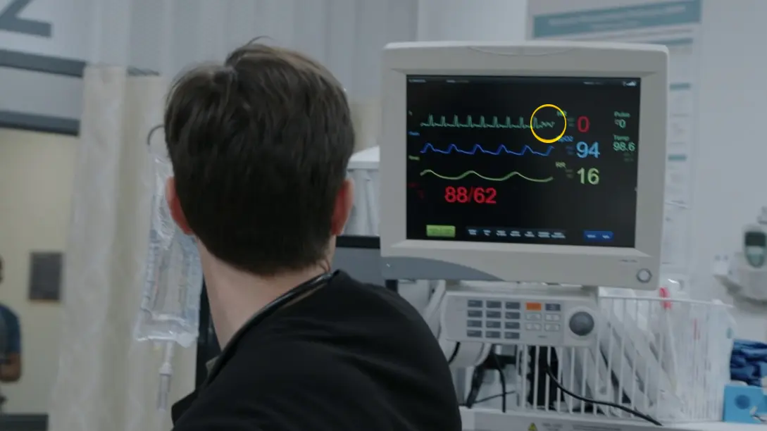
Patient presents with acute coronary syndrome symptoms and an EKG showing anterior STEMI, rapidly deteriorating into a witnessed Ventricular Fibrillation (V-Fib) cardiac arrest while awaiting Cath Lab transport.
+4
Anterior STEMI Arrival
Patient presents with acute coronary syndrome symptoms and an EKG showing anterior STEMI, rapidly deteriorating into a witnessed Ventricular Fibrillation (V-Fib) cardiac arrest while awaiting Cath Lab transport.
Medical Decision Making
The patient presents with a high-risk anterior STEMI. Initial medical management focuses on inhibiting platelet aggregation and reducing myocardial oxygen demand via aspirin and nitroglycerin. Anterior STEMIs carry a significant risk of lethal arrhythmias due to left ventricular ischemia. The patient suddenly loses consciousness and loses radial pulses, with the monitor demonstrating Ventricular Fibrillation. This necessitates an immediate pivot from medical management to the ACLS algorithm: initiating high-quality chest compressions, delivering early unsynchronized defibrillation, and establishing airway protection with high-flow oxygen.
Diagnostics & Findings
- 12-Lead EKG (performed at Triage, showing Anterior STEMI)
- Continuous cardiac monitoring
- Physical exam (No murmurs noted, radial pulses initially symmetric, then rapidly lost)
Findings:
- Active substernal chest pain 'elephant on my chest' and diaphoresis
- Sudden loss of consciousness and loss of pulses
- Monitor confirms Ventricular Fibrillation (V-Fib)
Interventions
- Administered 324 mg chewable Aspirin
- Administered Sublingual Nitroglycerin (0.4 mg x 3 doses)
- Initiated immediate manual chest compressions (Whitaker)
- Defibrillator charged and delivered unsynchronized shock at 200 Joules
- Initiated Bag-Valve-Mask (BVM) ventilation on 100% FiO2
- Prepared equipment for endotracheal intubation
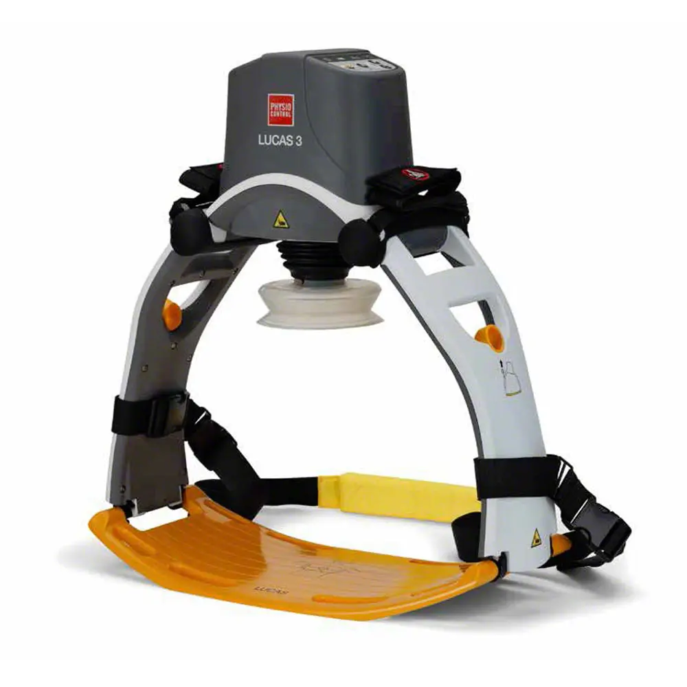
- Transitioned from manual CPR to mechanical CPR device (LUCAS)
⮑ Outcome & Reassessment
Patient did not convert after the initial 200J shock and remained in pulseless V-fib. Mechanical compressions (LUCAS) were started as resuscitation continued.
Clinical Media
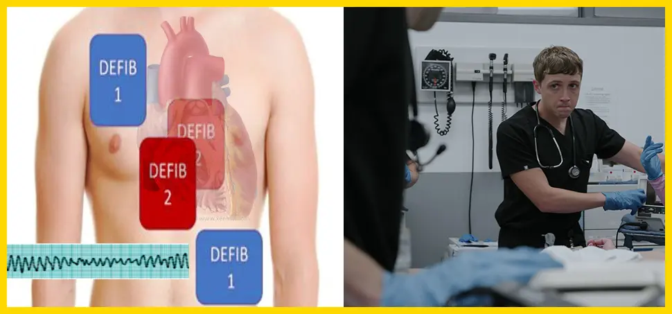
Double Sequential Defibrillation (DSED)
Failure of standard ACLS defibrillation to convert the V-fib rhythm.
+1
Double Sequential Defibrillation (DSED)
Failure of standard ACLS defibrillation to convert the V-fib rhythm.
Medical Decision Making
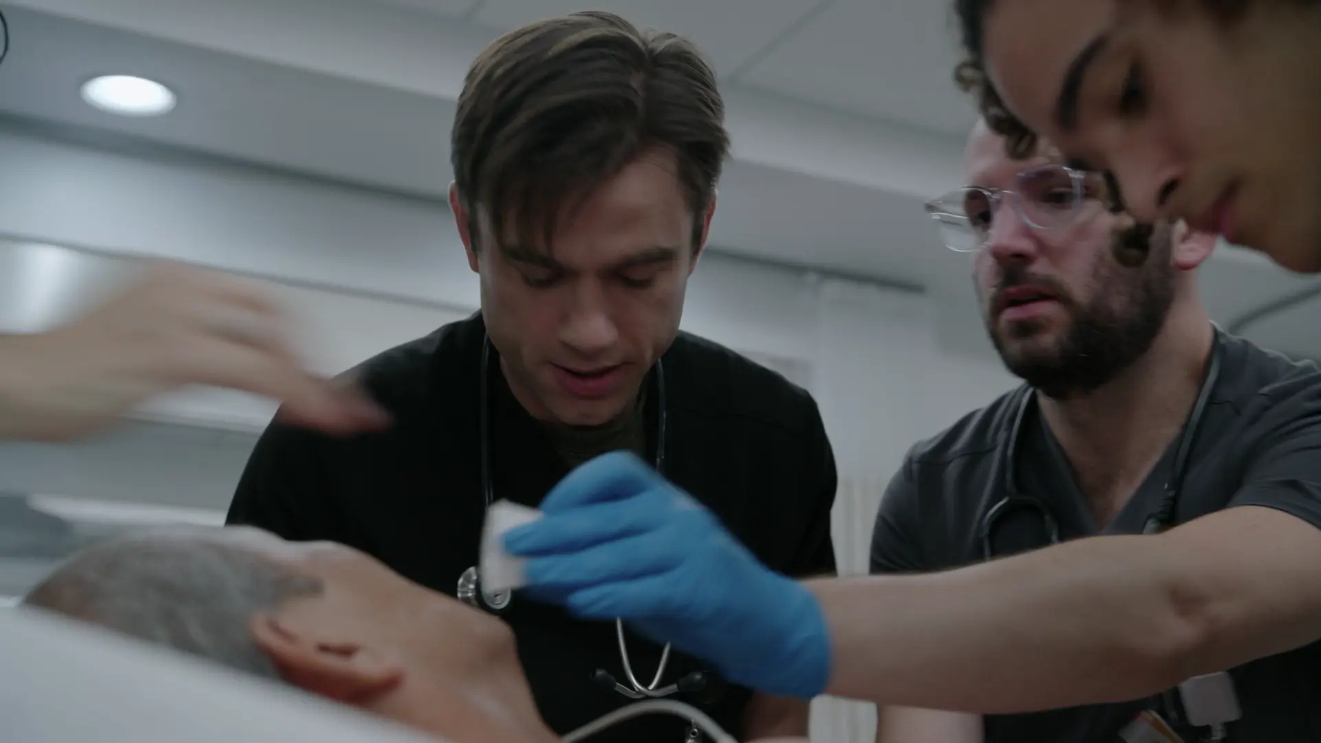
Standard defibrillation and epinephrine are failing. Will attempt Double Sequential Defibrillation (DSED), theorizing that a first shock lowers the defibrillation threshold and a rapidly subsequent second shock from a different vector can convert stubborn fibrillating myocytes. Also establishing an arterial line for real-time blood pressure monitoring and ABG access.
Diagnostics & Findings
- Arterial line placement
Findings:
- Persistent V-Fib
- No pulse on A-line
Interventions
- Epinephrine 1mg IV pushed
- Amiodarone requested
- Double Sequential Defibrillation setup (Right sternal border, Apex)
- Two 200J shocks delivered sequentially
⮑ Outcome & Reassessment
No change. Patient remains in refractory V-fib, pulseless.
Clinical Media
VA-ECMO Cannulation
DSED failure leaves eCPR as the final salvage option for an otherwise viable patient with a reversible cause (coronary occlusion).
+1
VA-ECMO Cannulation
DSED failure leaves eCPR as the final salvage option for an otherwise viable patient with a reversible cause (coronary occlusion).
Medical Decision Making
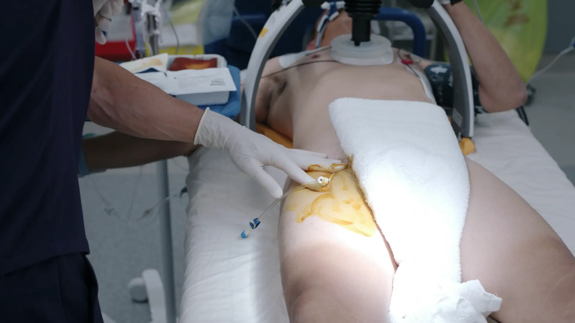
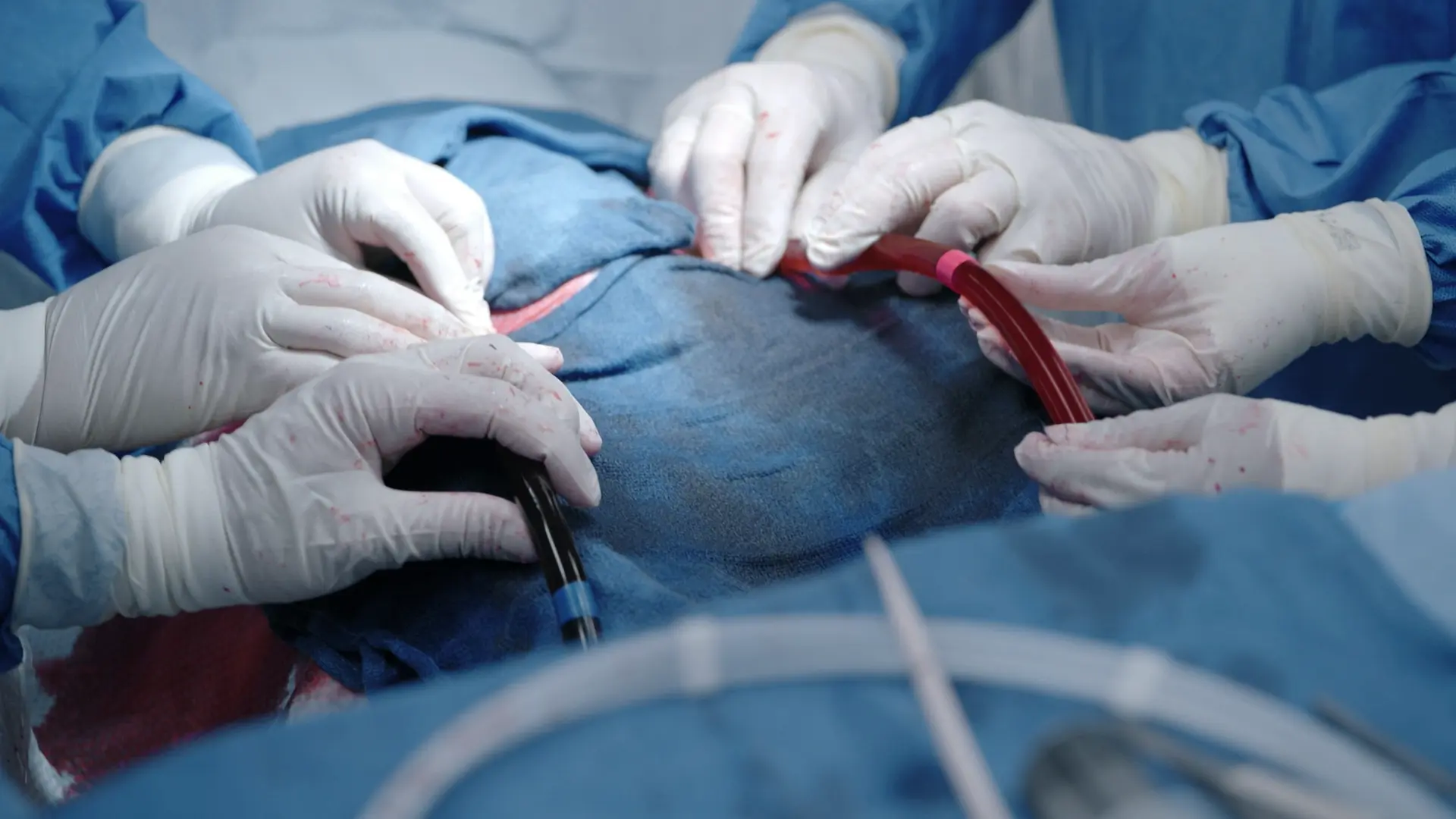
Patient meets criteria for Extracorporeal Cardiopulmonary Resuscitation (eCPR):witnessed in-hospital arrest with immediate CPR (essentially no no-flow time), an initial shockable rhythm, an easily reversible cause (STEMI requiring cath), and no obvious terminal comorbidity. VA-ECMO will bypass the heart and lungs, perfusing the body so the patient can survive transport to the Cath Lab. a cannula is a thin, flexible tube inserted into a body cavity, or blood vessel for medical treatment. it is fundamentally used to deliver or remove fluids, administer medications, or supply oxygen. Cannulation is a sterile surgical procedure because bacteria introduced into the circuit can cause catastrophic infection. a 'garden hose' drains blood from the right femoral vein to the ECMO machine for oxygenation, and a second hose returns it into the left femoral artery, so the machine does the work of both heart and lungs. An IJ central line is requested because the ECMO team will want central access for infusions and monitoring.
Diagnostics & Findings
- ABG drawn (looks good/acceptable for continuation)
Findings:
- Cannulation started
Interventions
- Sterile procedural setup
- 7-French Cordis introducer placed
- Right femoral vein (for venous drainage)
- Left femoral artery (for oxygenated blood return)
- Internal Jugular (IJ) line placed at neck
⮑ Outcome & Reassessment
Access is obtained without complication on screen; CPR continues throughout and resuscitation stays in the ECMO team's hands.
Clinical Media
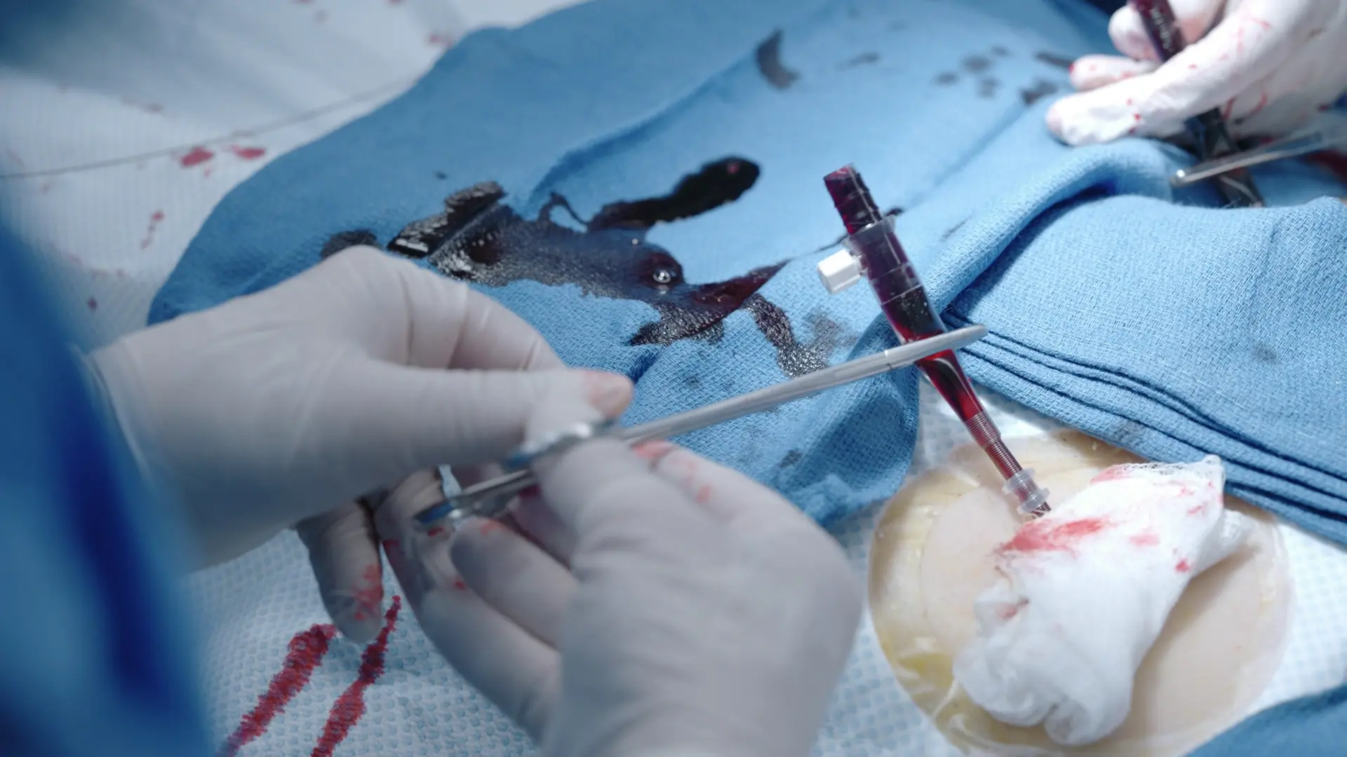
First Cannula Placement
Cannulas are being placed and Whitaker asks why ECMO was not tried on a prior patient (Mr. Milton, the gallbladder patient).
+1
First Cannula Placement
Cannulas are being placed and Whitaker asks why ECMO was not tried on a prior patient (Mr. Milton, the gallbladder patient).
Medical Decision Making
The vessel is serially dilated until it accepts the largest cannula (Seldinger technique), and the first line is placed and clamped to prevent air entry or blood loss. The teaching point is the eCPR criteria as Dr. Langdon states them: CPR started within about 5 minutes and an initial rhythm of V-fib, V-tach or PEA. Mr. Milton met neither, so he was not a candidate, whereas Mr. Chang had a witnessed arrest with immediate CPR and an initial rhythm of VF.
Diagnostics & Findings
Findings:
- Serial dilation until the largest cannula fits
- First cannula in and clamped
Interventions
- First ECMO cannula placed and clamped
⮑ Outcome & Reassessment
Cannulation proceeding; the second cannula is still needed. No change in rhythm.
Clinical Media
Initiation of VA-ECMO
Both cannulas are placed; the circuit must be connected and started to restore perfusion.
+2
Initiation of VA-ECMO
Both cannulas are placed; the circuit must be connected and started to restore perfusion.
Medical Decision Making
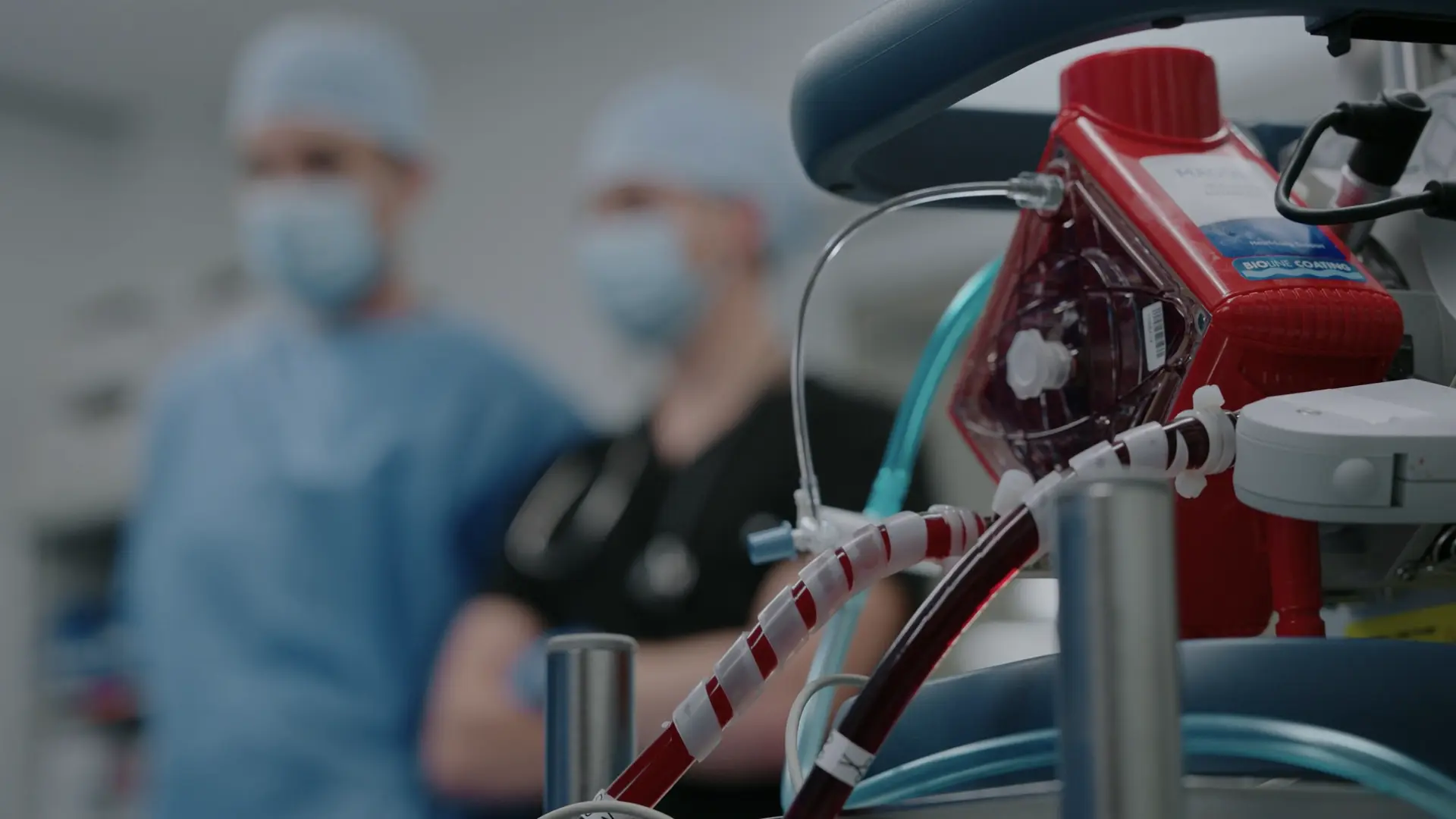
The two catheters are connected to the circuit lines with strict attention to air bubbles. Clamps are released and flow is established, mechanical CPR is stopped. A MAP of 74 confirms adequate perfusion pressure on circuit flow alone. ECMO has taken over heart and lung function and once the cath team opens the blocked artery the heart can be restarted and the patient could recover well.
Diagnostics & Findings
- MAP monitoring
Findings:
- MAP 74 on ECMO
- Circuit flowing without air bubbles
Interventions
- Cannulas connected to ECMO lines
- Circuit started and clamps released
- LUCAS stopped
- VA-ECMO established
⮑ Outcome & Reassessment
Excellent perfusion with MAP 74. Patient is stabilized on ECMO and awaits catheterization.
Clinical Media
Diagnoses & Disposition
Evolving Diagnoses
- [S01E07]Acute anterior ST-elevation myocardial infarction (STEMI)
- [S01E07]Ventricular fibrillation cardiac arrest complicating anterior STEMI
- [S01E07]Refractory ventricular fibrillation after epinephrine, amiodarone and double sequential defibrillation
- [S01E07]Cardiac arrest bridged with peripheral VA-ECMO (ECPR) pending revascularization
Current Disposition
Stabilized on VA-ECMO with a MAP of 74; transferring to Cardiology/Cath Lab for emergent percutaneous coronary intervention (PCI).
Casebook Analysis
Episode Context
This case highlights cutting-edge emergency resuscitation protocols and functions as a high-stakes, adrenaline-fueled set piece for the medical team. It juxtaposes the patient's calm, fatalistic acceptance of his potential death 'today is the day I'll be joining her' with the medical team's relentless, multi-modal fight to keep him alive. It also serves as a teaching moment for Whitaker.
Attending's Review
Medical Accuracy
The medical depiction is exceptionally accurate. The progression from an acute anterior STEMI into sudden V-fib is a classic and lethal presentation. The team's use of Double Sequential Defibrillation (DSED) correctly reflects modern evidence-based salvage therapies for refractory V-fib. Furthermore, the explanation and anatomical execution of VA-ECMO (eCPR) draining venous blood from the right femoral vein, oxygenating it, and pumping it back into the left femoral artery is technically precise. The criteria mentioned for ECMO (CPR within 5 minutes, shockable rhythm) align perfectly with real-world eCPR protocols.
Complications & Errors
- Langdon's summary that opening the artery will 'restart' the heart and leave him 'basically good as new' overstates the outlook: reperfusion arrhythmias, myocardial stunning, bleeding, limb ischemia and neurologic injury are all still real risks on ECMO.
Clinical Pearls
In acute coronary syndrome, aspirin should be chewed rather than swallowed whole. Chewing physically breaks down the tablet and bypasses the enteric coating delay, allowing for rapid buccal and gastrointestinal absorption to quickly inhibit platelet aggregation via COX-1 blockade.
Sublingual nitroglycerin relieves ischemic chest pain by promoting systemic venodilation, which decreases venous return (preload) and subsequently reduces myocardial oxygen demand. While generally safe in anterior STEMIs, it is contraindicated in inferior STEMIs with right ventricular involvement, as those patients are highly preload-dependent and nitroglycerin can precipitate profound, life-threatening hypotension.
Double Sequential Defibrillation (DSED) involves delivering two shocks rapidly from two different vectors (e.g., anterolateral and anteroposterior). It lowers the defibrillation threshold and redirects energy to target fibrillating myocytes unaffected by standard pad placement.
ECMO stands for Extracorporeal Membrane Oxygenation. The specific modality used for cardiac arrest is VA-ECMO (Veno-Arterial ECMO), which provides both hemodynamic (cardiac) and respiratory support by draining venous blood, oxygenating it extracorporeally, and pumping it directly into the arterial system under pressure. This is distinct from VV-ECMO (Veno-Venous), which provides only respiratory support.
ECMO is classified into two primary therapeutic modalities: VA-ECMO (Veno-Arterial) and VV-ECMO (Veno-Venous). eCPR (Extracorporeal CPR) strictly utilizes VA-ECMO, which drains deoxygenated venous blood often via the femoral vein, oxygenates it, and pumps it directly into the arterial system via the femoral artery. This provides both complete hemodynamic (cardiac) and respiratory support, bypassing the arrested heart. Conversely, VV-ECMO returns oxygenated blood to the venous system, providing only respiratory support and thereby requiring a functioning native heart to pump the blood systemically, VV-ECMO can be indicated in Severe ARDS, severe pneumonia, severe asthma.
eCPR (VA-ECMO for cardiac arrest) is a highly effective salvage therapy but requires strict inclusion criteria: witnessed arrest, immediate bystander/medical CPR, an initial shockable rhythm (VT/VF), and a reversible etiology such as a myocardial infarction.
In Veno-Arterial VA-ECMO, blood is drained from a large vein (commonly the femoral vein), passed through an oxygenator, and returned under pressure to a large artery (commonly the femoral artery), entirely bypassing the native heart and lungs.
Similar Cases from The Show
Ed Gellin
Code STEMI

Nandi Jawaharlal
The Beauty Influencer
Terrence
Dr. Google
Related Specialties
Chart Navigation
Harvey Chang
C/OSubsternal chest pain with diaphoresis (10 minutes duration)